
Researchers find how the intestinal wall folds by measuring forces
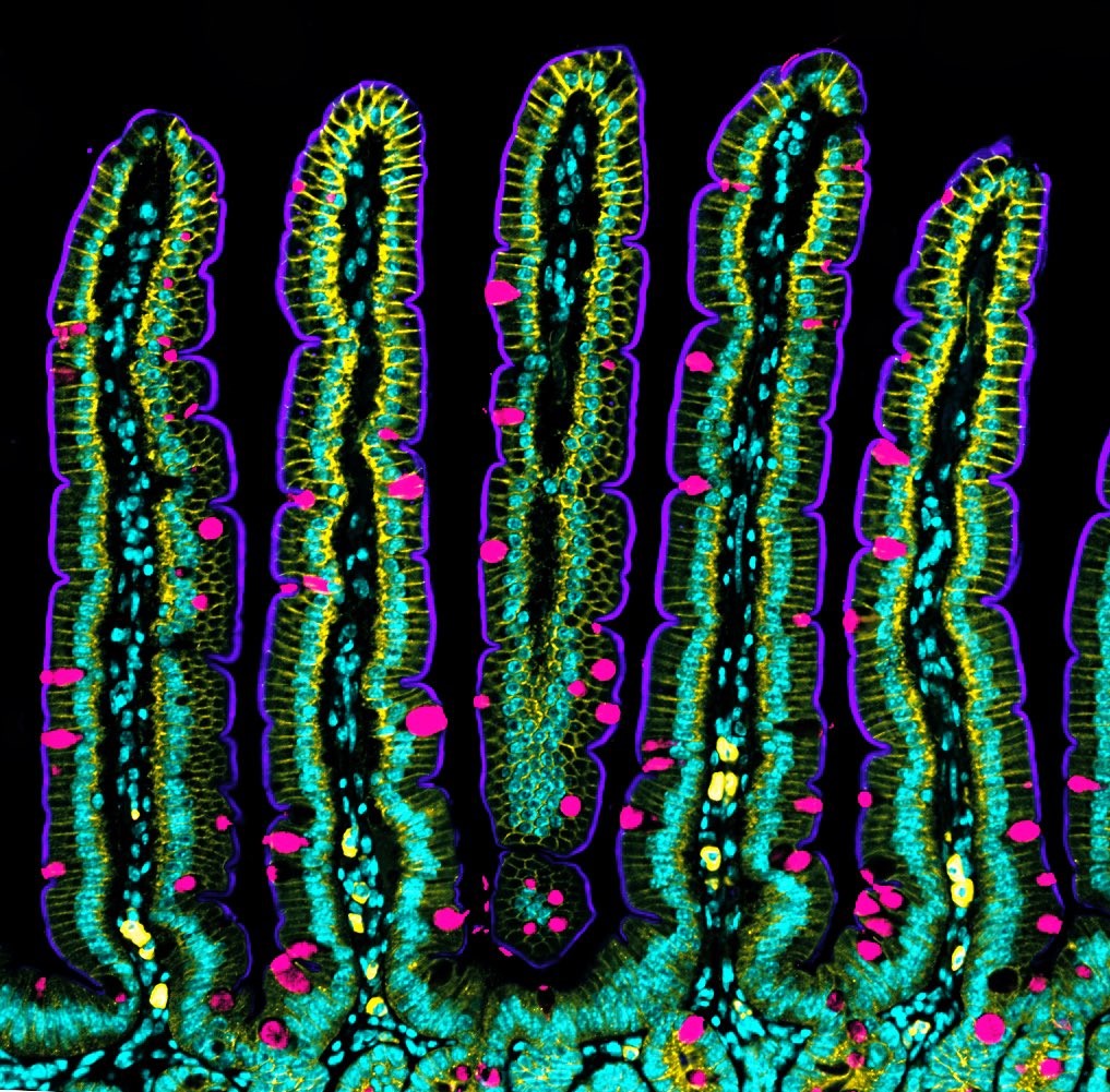
The human intestine is formed by more than 40 square meters of tissue, with many folds in the internal surface that remind of valleys and mountain peaks. Among other functions, these folds enable an increase in nutrient absorption. Also, the intestine is under a constant renewal, which involves that, approximately every five days, all cells of its internal wall are renewed to guarantee a proper intestinal functioning. To date, it was known that such renewal was possible thanks to the stem cells that are protected in the intestinal crypts, and which give rise to new differenced cells. However, the process that leads to the concave shape of crypts and the migration of the new cells to the intestinal top was still an enigma.
The human intestine is formed by more than 40 square meters of tissue, with many folds in the internal surface that remind of valleys and mountain peaks. Among other functions, these folds enable an increase in nutrient absorption. Also, the intestine is under a constant renewal, which involves that, approximately every five days, all cells of its internal wall are renewed to guarantee a proper intestinal functioning. To date, it was known that such renewal was possible thanks to the stem cells that are protected in the intestinal crypts, and which give rise to new differenced cells. However, the process that leads to the concave shape of crypts and the migration of the new cells to the intestinal top was still an enigma.
Now, an international team led by Xavier Trepat, researcher at the Department of Biomedicine of the UB and ICREA research professor at the Institute of Bioengineering of Catalonia (IBEC), has dismantled the mechanism with which the crypts adopt and maintain the concave shape and how the cell migration towards the top occurs without the intestine losing its characteristic folding form. The study, published in the prestigious journal Nature Cell Biology, shows that this process is possible thanks to mechanical forces of the cells. To confirm it, researchers combined the computational modelling with mice cellular intestinal organoids.
By using mice stem cells and bioengineering and mechanobiology techniques, researchers worked on mini-intestines, organoids that reproduce the three-dimensional structure of valleys and peaks of the tissue in vivo, and its functions. Using microscopy technologies developed in the same group, for the first time, they carried out a series of high-resolution experiments that enabled them to obtain 3D maps showing the used forces by each cell. Moreover, with this in vitro model, the researchers showed that the movement of the new cells towards the top is controlled by mechanical forces of the same cells, specifically the cytoskeleton, a network of strands that determines and keeps the cell form.
Reference article:
C. Pérez-González, G. Ceada, F. Greco, M. Matejčić, M. Gómez-González, N. Castro, A. Menéndez, S. Kale, D. Krndija, A. G. Clark, V. Ram Gannavarapu, A. Álvarez-Varela, P. Roca-Cusachs, E. Batlle, D. Matic Vignjevic, M. Arroyo and X. Trepat. "Mechanical compartmentalization of the intestinal organoid enables crypt folding and collective cell migration". Nature Cell Biology, June 2021. DOI: https://doi.org/10.1038/s41556-021-00699-6